Ch. 14
NUCLEIC ACIDS
Function: Nucleic Acids carry hereditary (genetic) information
for all cells
RNA (Ribonucleic Acid)
Nucleotides are the monomers (subunits) of all nucleic acids.
a) Phosphate group (PO4)
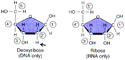
b) Five carbon sugar: deoxyribose- 1 less oxygen than ribose
(RNA)
c) Nitrogen base
Purines: adenine, guanine (double
rings)
Pyrimidines: thymine, cytosine
(uracil-RNA only) (single rings)
 Diagram at
right is a simplified model of a nucleotide.
Diagram at
right is a simplified model of a nucleotide.
Sugar structure
A prime (') notes where a carbon is located on the sugar
Phosphate attaches to the 5' carbon
Nitrogen bases attach to the 1' carbon
-OH attaches to 3' carbon ( as well as the phosphate of the
next nucleotide.
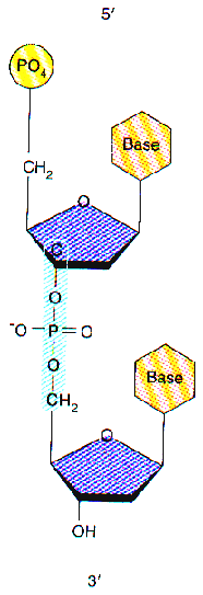 Nucleic
Acid Chain Structure
Nucleic
Acid Chain Structure
Phosphate at 5', hydroxyl at 3' form chains
Dehydration synthesis links 3' to 5' ends (Hydroxyl 3' to phosphate
5') 5'xxxxx3' - 5'xxxxx3' - 5'xxxxx3' etc.
Double helix structure
Double helix describes the spiral, double-stranded shape of DNA.
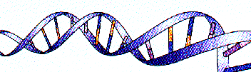
Bases point inward
Large purine (2 rings) always paired with small pyrimidine
(1 ring) for consistent width
Adenine - thymine for two bonds (A-T)
Guanine - cytosine form three bonds (C-G)
Antiparallel: DNA is antiparallel. Its two strands run in opposite
directions.
Fraenkel - Conrat Experiment : tobacco mosaic virus ( TMV ) and
rib grass virus ( HRV ) (Read pp. 282-83)
Retro viruses : ( retro = backwards
) RNA virus uses RNA to code DNA of host ( HIV causing AIDS )
Reverse transcriptase enzyme codes DNA from virus RNA, inserts
it into host cell.
Slichter